
BMV — Opening a Narrowed Mitral Valve Without Surgery
A catheter-based procedure for patients with mitral stenosis. BMV uses a balloon to separate fused valve leaflets and restore blood flow — without open-heart surgery.




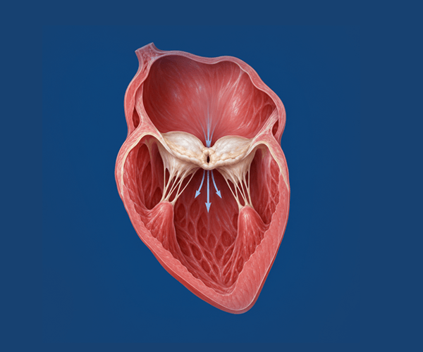
What is mitral stenosis?
Mitral stenosis is a narrowing of the mitral valve that restricts blood flow from the left atrium to the left ventricle. In India, it is most commonly caused by rheumatic heart disease.
As the narrowing progresses, the heart has to work harder to push blood through the restricted opening. Over time, this can cause symptoms — and if untreated, lead to serious complications including atrial fibrillation and heart failure.
Common symptoms
- Breathlessness — on exertion, or when lying flat
- Fatigue and reduced exercise tolerance
- Palpitations, which may indicate atrial fibrillation
- Swelling of the legs or ankles
- In more advanced disease: coughing up blood (haemoptysis)
A proper medical evaluation — including echocardiography — is needed to confirm the diagnosis and determine severity.
What is Balloon Mitral Valvotomy?
BMV (also called PTMC — Percutaneous Transvenous Mitral Commissurotomy) is a catheter-based procedure to widen a narrowed mitral valve without open-heart surgery.
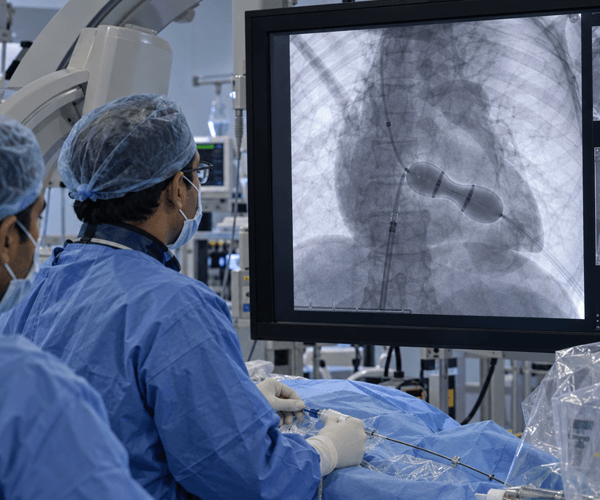
A thin, flexible catheter is advanced from the groin to the heart. A balloon at the tip of the catheter is positioned across the narrowed valve and inflated to separate the fused valve leaflets. This improves blood flow through the valve immediately.
Success rates apply to carefully selected patients with suitable valve anatomy. Individual outcomes vary. Source: ACC/AHA Valvular Heart Disease Guidelines; published BMV outcome data.
Who may be suitable for BMV?
BMV works best in patients whose valve leaflets are still flexible and relatively free of calcium. A detailed echocardiogram — and sometimes a transoesophageal echo — is used to assess suitability.






BMV is not suitable for every patient with mitral stenosis. Each case is reviewed by the Heart Team using clinical information and imaging to determine the most appropriate treatment — whether BMV, surgical commissurotomy, or mitral valve replacement.
How BMV is performed
BMV is carried out in a cardiac catheterisation laboratory under fluoroscopic and echocardiographic guidance. It is a planned procedure, typically performed under conscious sedation or light general anaesthesia.




What outcomes does BMV achieve?
In appropriately selected patients, BMV delivers meaningful improvement in valve function and symptoms. The data below reflects outcomes at specialist centres in carefully selected patients — individual results vary.
Sources: Published BMV/PTMC outcome data; ACC/AHA Guidelines for the Management of Patients with Valvular Heart Disease; Iung B et al., European Heart Journal. These figures represent outcomes in selected patient cohorts and should not be interpreted as individual expected outcomes. All patients are assessed individually by the Heart Team before any recommendation is made.

Recovery after BMV
Recovery is generally faster than after open-heart surgery. The exact timeline depends on each patient's overall health and how the heart responds.
In hospital (typically 1–3 days)
- Monitoring of heart rhythm, blood pressure and access site
- Echocardiogram before discharge to assess valve result
- Most patients begin walking within 24 hours of the procedure
- Anticoagulation is reviewed — may continue if atrial fibrillation is present
After discharge
- Return to light daily activities within 1–2 weeks for most patients
- Follow-up echocardiogram at 1 month, then annually
- Medical management continues — diuretics, rate control, anticoagulation as needed
- Regular cardiac review is important: BMV may not be permanent in all patients
Risks and transparency
As with all cardiac catheter procedures, BMV carries risk. The exact risks depend on individual patient health, valve anatomy and the clinical context. All risks are discussed with patients before proceeding.
Procedure-related risks
- Worsening of mitral valve leakage (mitral regurgitation) — most common significant complication
- Cardiac tamponade (fluid around the heart) — rare
- Stroke or embolism — reduced by TEE screening before the procedure
- Need for emergency cardiac surgery
- Access-site complications (bleeding, bruising)
Longer-term considerations
- Restenosis — the valve may narrow again over time; some patients need a repeat procedure or surgery after 5–15 years
- Residual atrial fibrillation — anticoagulation may be needed long-term
- Ongoing follow-up is important to monitor valve function and symptom status
Frequently asked questions
BMV (Balloon Mitral Valvotomy) opens the narrowed valve using a balloon through a catheter — no chest incision or heart-lung bypass is required. Surgical commissurotomy and mitral valve replacement are also effective treatments. In suitable patients, BMV achieves comparable outcomes to surgical commissurotomy with a shorter recovery. The most appropriate approach depends on valve anatomy, severity and each patient's overall health.
BMV is not necessarily a permanent solution. In well-selected patients with pliable valves and a good procedural result, many remain free from restenosis for 10–15 years. However, some patients (approximately 35–50% by 5–10 years) will eventually need a repeat procedure or surgery as the disease progresses. Regular echocardiographic follow-up is important.
Patients with heavily calcified, rigid valves, significant mitral regurgitation, or a clot in the left atrium are generally not suitable for BMV. In these cases, surgical options — valve repair or replacement — may be more appropriate. Each case is assessed individually by the Heart Team using echocardiography and clinical information.
Assessment typically includes a transthoracic echocardiogram and a transoesophageal echocardiogram (TOE/TEE) — which is essential to check for clots in the left atrium and to assess valve anatomy in detail. Blood tests and other routine investigations are also performed. The Wilkins scoring system helps determine how suitable the valve is for BMV.
The procedure typically takes 1–2 hours, though this varies depending on anatomy and procedural complexity. Patients spend additional time in the catheterisation lab for preparation and monitoring after the procedure. Most patients are transferred to a ward within a few hours of completing the procedure.
Diagnosed with mitral stenosis?
Understand whether BMV is appropriate for your condition. Our Heart Team can review your echo reports and explain your options.