
TMVR — Replacing a Mitral Valve Without Open-Chest Surgery
For patients with complex mitral valve disease — particularly failed previous valve surgery — TMVR offers a catheter-based route to valve replacement when conventional surgery carries high risk or is not feasible.
What is TMVR?
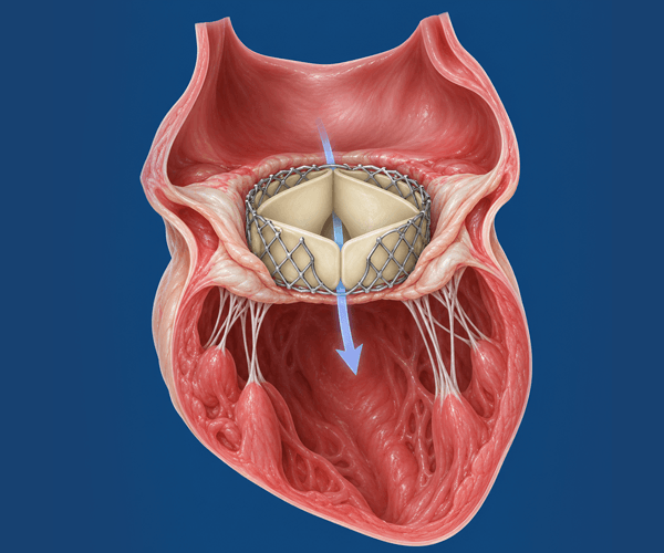
TMVR (Transcatheter Mitral Valve Replacement) is a catheter-based procedure in which a prosthetic valve is deployed inside a diseased mitral valve — without opening the chest.
Unlike TEER (MitraClip), which clips and repairs the existing valve, TMVR replaces the valve entirely. This is indicated in patients where the mitral valve cannot be effectively repaired, or where a previous repair or bioprosthetic valve has failed.
How is TMVR different from TEER?
Types of TMVR
TMVR is used in different clinical situations. The most established use is valve-in-valve replacement for failed bioprosthetic valves. Native valve TMVR is a newer, evolving area.

- Growing evidence base
- Avoids complex redo cardiac surgery
- Particularly relevant in elderly or frail patients

- Suitable when surgical repair has failed
- Ring provides anchoring structure for the device
- Case-by-case anatomical assessment required

- Selected high-risk patients only
- Requires complex anatomical planning (CT, echo)
- Evidence continues to develop — discussed case-by-case
Who may be considered for TMVR?
TMVR is a complex procedure reserved for specific clinical situations. Detailed pre-procedural imaging — 3D echocardiography and CT — is essential before any decision is made.






TMVR is not a routine procedure. Each case must be individually reviewed by a Heart Team including structural cardiologists, cardiac surgeons, and imaging specialists before any recommendation can be made.
How TMVR is performed
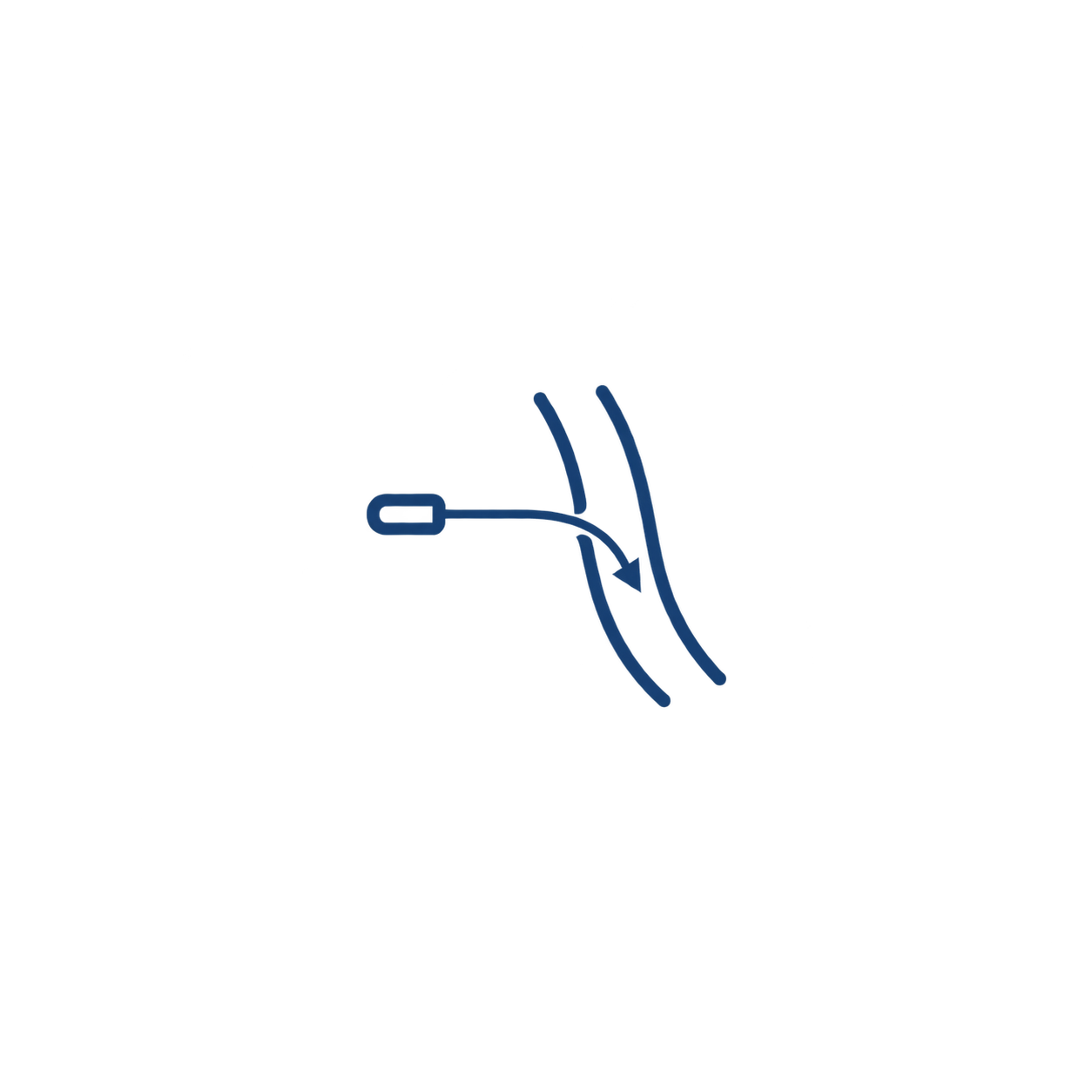
TMVR is performed in a specialised cardiac catheterisation laboratory or hybrid operating theatre. Advanced 3D echocardiography, CT planning and fluoroscopy are central to the procedure.



What does the evidence show?
TMVR evidence differs significantly between its applications. Valve-in-valve TMVR for failed bioprosthetics has a growing evidence base. Native valve TMVR remains an evolving field with more limited data.
Valve-in-Valve and Valve-in-Ring — key evidence points
- Published registry data (VIVID registry) supports ViV TMVR for failed surgical bioprostheses in high-risk patients as an established alternative to redo surgery
- Technical success rates of 90–95% reported at experienced centres for ViV cases
- 30-day mortality varies by patient risk profile — registry data reports approximately 4–7% in high-risk populations
- LVOT obstruction remains a key risk specific to TMVR, occurring in 2–10% of cases depending on anatomy — CT pre-screening is essential to identify this risk
- Valve-in-ring outcomes are generally good but require careful anatomical assessment; ring type affects anchoring and haemodynamics
Sources: VIVID Registry; published TMVR outcome data; ACC/AHA Valvular Heart Disease Guidelines. Evidence is evolving — outcomes depend heavily on patient selection, anatomy and centre experience. These figures represent registry populations, not individual expected outcomes. Every TMVR case is reviewed individually by the Heart Team before any recommendation.
Recovery after TMVR
Recovery varies significantly depending on the access route used, the patient's overall health, and the clinical context. There is no fixed recovery timeline that applies to all TMVR patients.
Transseptal approach (more common)
- Hospital stay: typically 3–5 days depending on clinical progress
- Most patients can begin mobilising within 1–2 days
- Shorter overall recovery than transapical or open surgical approaches
Transapical approach (when required)
- A small chest incision is needed — slightly longer recovery than transseptal
- Hospital stay: typically 4–7 days
- Pain management and respiratory physiotherapy important in early recovery
Follow-up after discharge
- Echocardiogram at 1 month, 6 months and 12 months to monitor valve function
- Anticoagulation is usually required — type and duration determined by the valve and clinical situation
- Regular cardiac review — TMVR patients require ongoing specialist monitoring

Risks and transparency
TMVR is a complex, high-risk procedure. The balance of risk and benefit depends on the individual patient's anatomy, prior history and overall health. All risks are discussed openly with patients and families before proceeding.
Procedure-related risks
- LVOT (left ventricular outflow tract) obstruction — a specific risk in TMVR, identified by CT pre-screening
- Paravalvular leak — leakage around the prosthetic valve
- Stroke or embolism
- Need for a permanent pacemaker
- Cardiac tamponade (fluid around the heart)
- Access site complications
- Need for conversion to open surgery
Longer-term considerations
- Valve durability — bioprosthetic valves have finite durability; a further procedure may be needed in the future
- Anticoagulation — lifelong anticoagulation therapy is often required
- Need for device-specific surveillance imaging
- The procedure does not address coronary artery disease or other underlying cardiac conditions
Frequently asked questions
TEER (Transcatheter Edge-to-Edge Repair, including MitraClip) clips and repairs the existing mitral valve leaflets. The native valve remains in place. TMVR replaces the mitral valve entirely with a prosthetic valve — it is used when the anatomy is too damaged or complex for repair, or when a previous surgical repair or bioprosthetic valve has failed. Both are catheter-based and avoid standard open-chest surgery.
It depends on the application. Valve-in-valve TMVR — replacing a failed bioprosthetic mitral valve — is the most established indication, supported by registry data and guideline recognition. Valve-in-ring TMVR is also performed at specialist centres. Native mitral valve TMVR remains an evolving field with more limited evidence, and is generally considered only in selected high-risk patients at experienced centres.
LVOT obstruction (left ventricular outflow tract obstruction) is a specific risk in TMVR where the prosthetic valve alters the geometry of the heart in a way that obstructs blood flow out through the aortic valve. It can be serious and, in some cases, fatal. It is not a risk with TEER. CT scanning before the procedure allows the Heart Team to identify patients at high LVOT risk and plan accordingly — including deciding whether TMVR is appropriate at all in certain anatomies.
In most cases, yes. Anticoagulation is typically required after TMVR — the type and duration depend on the prosthetic valve used, the patient's cardiac rhythm and other clinical factors. The treating team will discuss the specific anticoagulation plan with you before and after the procedure.
Suitability cannot be determined without a detailed clinical assessment, echocardiogram, and CT scan. The Heart Team reviews all of these together. Key factors include the underlying mitral valve pathology, whether previous surgery has been performed, the anatomy of the valve and surrounding structures, and the patient's overall surgical risk. Contact our team to have your reports reviewed.
Complex mitral valve disease? Let us review your case.
TMVR may be an option when other treatments are not suitable. Our Heart Team can review your echo and CT reports to advise whether it applies to your situation.